
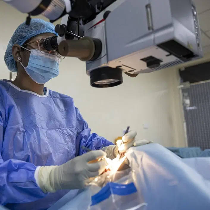
The Cornea Needs to Stay Clear to Provide Useful Vision
The cornea forms the clear front window of the eye. Light must pass through it before reaching the deeper structures responsible for vision. If the cornea becomes cloudy, scarred, swollen or significantly distorted, incoming light may no longer be focused properly.
The effect on vision depends on the location and severity of the damage. A small peripheral scar may cause relatively little difficulty, while a dense central scar can interfere significantly with visual clarity. Some corneal diseases mainly affect shape, while others damage the innermost endothelial cells responsible for maintaining normal corneal transparency.
Corneal transplantation becomes relevant when the damaged tissue cannot provide adequate vision and less invasive options are not expected to achieve the required improvement.
Who May Need a Corneal Transplant?
Corneal transplantation is generally considered after a detailed examination establishes that the problem originates from the cornea and that replacing damaged tissue offers a reasonable chance of improving or preserving vision. The underlying diagnosis strongly influences which transplant technique is appropriate.
Situations that may lead to consideration of transplantation include:
Advanced corneal scarring that blocks or distorts vision and cannot be adequately managed through other treatment.
Endothelial disorders that cause persistent corneal swelling because the innermost cell layer no longer functions effectively.
Selected advanced cases of keratoconus where the cornea has become severely irregular or scarred and other treatments are no longer sufficient.
Significant corneal injury or previous infection resulting in permanent loss of transparency.
Certain inherited corneal dystrophies that progressively affect corneal clarity or function.
Failure of a previous corneal graft when another transplant is considered appropriate.
Structural corneal damage severe enough that transplantation is needed to preserve the integrity of the eye.
Selected postoperative corneal problems in which endothelial or full-thickness replacement offers an appropriate treatment option.
A diagnosis such as keratoconus does not automatically mean transplantation is necessary. Earlier disease may be managed with glasses, contact lenses or treatments intended to stabilize the cornea. Surgery is considered according to disease severity, scarring, vision and individual anatomy.
Full-Thickness Transplantation Replaces the Entire Cornea
A penetrating keratoplasty, often shortened to PK, replaces the full thickness of the central cornea with donor tissue. It can be useful when disease or scarring affects multiple corneal layers rather than one isolated region.
During surgery, a circular portion of the patient's damaged cornea is removed and replaced with a corresponding donor graft. Sutures are typically required to hold the transplanted tissue in position while healing takes place.
Because the entire thickness of the cornea is replaced, recovery can be lengthy. Vision may change as the graft heals and as sutures influence corneal curvature. Full visual stabilization can take many months, and in some cases complete recovery may extend toward a year.
Full-thickness transplantation remains valuable, but it is no longer necessary for every corneal disease.
Partial-Thickness Surgery Preserves Healthy Corneal Layers
When only one portion of the cornea is diseased, surgeons may be able to replace that layer while preserving the patient's healthy tissue. These procedures are broadly known as lamellar keratoplasty.
This approach is important because corneal diseases do not always affect the entire structure. A patient with damaged inner endothelial cells may still have healthy outer corneal tissue. Another person with a structural disorder affecting the front and middle layers may have a healthy endothelium.
Choosing the transplant according to the affected layer can reduce unnecessary tissue replacement and, in several situations, lower particular complications compared with replacing the full corneal thickness.
DALK Can Be Considered When the Endothelium Is Healthy
Deep anterior lamellar keratoplasty, or DALK, replaces the front and deeper stromal portions of the cornea while preserving the patient's own endothelium.
This can be relevant in selected corneal disorders where the structural layers are damaged but the endothelial layer remains healthy. Because the patient's own endothelium is preserved, one important source of endothelial rejection is avoided.
Recent evidence comparing DALK with full-thickness transplantation has found broadly comparable visual outcomes in studied groups, while full-thickness transplantation was associated with a higher rate of rejection episodes and several complications. The trade-offs vary according to diagnosis and surgical complexity, so DALK is not automatically the better choice for every patient.
DMEK Replaces an Extremely Thin Inner Layer
For diseases primarily affecting the corneal endothelium, Descemet membrane endothelial keratoplasty, or DMEK, can replace only the innermost diseased tissue.
The donor graft used in DMEK is extremely thin. Preserving most of the patient's original cornea can allow relatively rapid visual recovery and good optical quality in suitable cases. DMEK has become increasingly important in the treatment of endothelial disease.
Because the graft is thin and delicate, postoperative positioning and attachment are particularly important. Sometimes the graft does not remain fully attached and an additional air or gas injection, commonly called rebubbling, may be needed.
Comparative evidence suggests that DMEK can provide better visual acuity than ultrathin DSAEK in appropriate endothelial cases, although DMEK tends to have a higher need for rebubbling.
DSAEK Uses a Slightly Thicker Donor Layer
Descemet stripping automated endothelial keratoplasty, or DSAEK, also treats endothelial dysfunction but transplants a somewhat thicker layer of donor tissue than DMEK.
Both procedures preserve the outer portion of the patient's cornea and avoid the large full-thickness wound associated with penetrating keratoplasty.
DMEK may offer faster or sharper visual recovery in some patients, while DSAEK can offer technical or postoperative advantages in selected situations. Neither name should be treated as a universal winner. The patient's eye, previous surgery, anatomy and the surgeon's experience influence which technique is more suitable.
Donor Tissue Is Only One Part of Successful Surgery
Corneal transplantation depends on healthy donor tissue, but graft quality alone does not determine the result.
The surgeon must choose the correct procedure, prepare the recipient cornea accurately and manage postoperative healing. The condition of the eye before surgery also matters. Severe inflammation, abnormal blood vessels growing into the cornea and previous graft rejection can increase the risk of immune complications.
For international patients, donor-tissue availability should be discussed before travel. A surgical date should not be assumed to guarantee that every type of donor graft can always be provided on exactly the desired schedule.
The treating team should be able to explain what tissue is required, how the operation is scheduled and what happens if the surgery needs to be postponed.
Corneal Transplant Surgery Is Usually Followed Closely
Corneal transplantation is commonly performed as an outpatient procedure. Depending on the individual situation, surgery may use local anaesthesia with medication for relaxation or general anaesthesia.
After the damaged corneal tissue has been removed or separated, donor tissue is positioned according to the chosen transplant technique. Full-thickness grafts generally require sutures, while endothelial techniques rely on different methods to secure a thin internal graft.
Follow-up begins quickly. A postoperative examination is typically required the following day so that graft position, eye pressure, wound condition and early healing can be assessed.
This early review is one reason an international patient should not plan to fly home immediately after surgery.
Vision Can Be Blurry During Early Recovery
Patients should not expect perfect vision when they leave the operating room.
Blurred vision, sensitivity and temporary visual fluctuation can occur during healing. The speed of visual improvement varies considerably according to the transplant type. Endothelial procedures may recover differently from full-thickness grafts, while penetrating keratoplasty can require a much longer period before refraction and corneal shape stabilize.
Patients may also need changes in glasses or other visual correction once the transplanted cornea has healed sufficiently.
The final outcome should therefore be judged over the appropriate surgical timeline rather than from an early postoperative vision test.
Eye Drops Are Part of the Transplant, Not an Optional Extra
Postoperative medication commonly includes prescribed eye drops designed to support healing and reduce inflammation and rejection risk. These medicines may need to continue for a prolonged period according to the transplant type and individual risk profile.
Stopping medication simply because the eye feels comfortable can be dangerous.
Patients should know which drops they are taking, how often they should be used and who is responsible for adjusting them after they return home. Long-term steroid drops can require monitoring because treatment itself may affect eye pressure in some patients.
A clear written medication plan is particularly important when postoperative care will eventually be transferred to an ophthalmologist in another country.
Corneal Rejection Can Happen After an Initially Successful Operation
The immune system can recognize transplanted corneal tissue as foreign and begin attacking the graft. This is known as corneal graft rejection. It can occur after the initial surgical recovery, which is why long-term awareness remains important.
Partial-thickness techniques generally have lower rejection risks than penetrating keratoplasty because less donor tissue is transplanted, although rejection is still possible with several lamellar procedures.
Rejection does not automatically mean that the graft is permanently lost. Prompt treatment can sometimes reverse an episode and preserve transplant clarity. Delay makes successful treatment less certain.
Which Symptoms Need Urgent Attention After a Corneal Transplant?
Most postoperative discomfort should gradually improve. Certain symptoms are different because they can indicate graft rejection or another complication and should be treated as reasons for prompt ophthalmic assessment.
Important warning signs include:
New or increasing eye pain, especially after the eye had previously been comfortable.
Increasing sensitivity to light that appears or becomes noticeably worse.
New redness of the operated eye, particularly when associated with visual change.
Cloudy or hazy vision after vision had begun to improve.
A noticeable deterioration in vision, rather than ordinary day-to-day fluctuation.
Increasing discharge or marked swelling, which may indicate infection or another postoperative problem.
A sudden change after trauma to the eye, even when the initial injury appears minor.
Any concerning symptom specifically identified by the operating surgeon as requiring urgent review.
A patient who has travelled home should not wait for the next trip to Turkey when these symptoms appear. Corneal rejection and infection are time-sensitive problems and should be assessed promptly where the patient currently is.
Other Complications Can Affect the Eye
Rejection is one of the best-known risks, but it is not the only possible complication.
Corneal transplant surgery can also be associated with infection, bleeding, increased eye pressure or glaucoma and retinal detachment. Depending on the transplant technique, graft detachment, irregular astigmatism or the need for additional procedures can also influence recovery.
Some grafts eventually fail despite treatment and may require another transplant. Risk is influenced by the underlying disease, previous eye surgery, inflammation, vascularization and the type of graft performed.
A realistic consultation should therefore discuss both the chance of visual improvement and the possibility of further treatment later.
Rubbing the Eye Can Be Risky During Healing
Protecting the operated eye is important after transplantation.
Patients are generally advised to avoid rubbing or pressing on the eye and may use glasses or an eye shield for protection during early recovery.
Physical activity restrictions depend on the type of surgery and stage of healing. A full-thickness graft secured with sutures presents different mechanical considerations from an endothelial transplant.
For this reason, advice about exercise, swimming, bending, lifting, contact sports and returning to work should come from the treating ophthalmologist rather than a standard online schedule.
Corneal Transplant Cost in Turkey Depends on the Technique
There is no single meaningful cornea transplant cost in Turkey because corneal transplantation includes several very different procedures.
A penetrating keratoplasty does not involve the same surgical process as DMEK, DSAEK or DALK. Donor-tissue preparation, operating-room requirements, anaesthesia, postoperative examinations and possible additional procedures can all affect the total treatment plan.
Patients should therefore ask whether a quotation includes donor tissue, surgery, anaesthesia, postoperative medication and scheduled follow-up.
A lower headline price has limited meaning when the exact transplant technique and follow-up plan are unclear.
What International Patients Should Verify Before Surgery
Corneal transplantation requires more continuity than many short-stay medical treatments. Turkey currently maintains lists of healthcare providers authorized to treat international health-tourism patients, with the current provider records dated July 28, 2026.
Current provider authorization is only one part of planning. Patients should know which ophthalmologist is responsible for the transplant, what technique is proposed and why, how donor tissue is arranged and how long they should remain locally for postoperative examinations.
It is also important to arrange ophthalmic follow-up at home before travelling whenever possible. A corneal graft may require medication adjustments and repeated examinations long after the initial operation.
Medical records should travel with the patient. The transplant type, surgical report, donor information available for clinical use, medication schedule and follow-up findings can all help another ophthalmologist continue care safely.
A Successful Corneal Transplant Is a Long-Term Partnership
Cornea transplant in Turkey can restore useful vision for selected patients with severe corneal disease, but success depends on much more than the surgical day itself.
Modern keratoplasty allows treatment to be tailored to the damaged layer. Penetrating keratoplasty replaces the full thickness of the cornea, while techniques such as DALK, DSAEK and DMEK preserve healthy tissue when only certain layers need replacement.
These options have different benefits, recovery patterns and complication profiles. DMEK, for example, can provide excellent visual outcomes in endothelial disease but has a greater likelihood of requiring rebubbling than some alternative endothelial techniques. DALK can avoid replacing healthy endothelium in appropriate anterior corneal disease.
The postoperative period is equally important. Patients need prescribed eye drops, eye protection and scheduled follow-up, while symptoms such as pain, increasing light sensitivity, redness or cloudy vision require prompt assessment because they can signal rejection.
For international patients, the best corneal transplant in Turkey is therefore not simply the procedure with the shortest stay or most attractive package. It is the operation that matches the exact corneal disease and comes with a realistic plan for donor tissue, early postoperative monitoring and long-term specialist care after returning home.
Note: This article is intended for general information and does not replace an individual ophthalmic examination. The need for corneal transplantation, choice of keratoplasty technique, donor-tissue requirements, medication and follow-up schedule should be determined by an eye specialist after detailed examination.